
Chronic Pain Diagnosis
- Chiropractor
- Physical therapist
- Neurosurgeon
- Pain medicine specialist
- Physiatrist
- Rheumatologist
- Orthopedic spine surgeon
Table of Contents
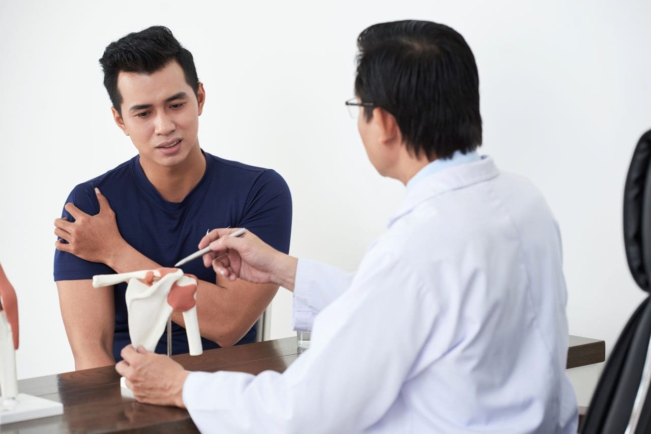
Chronic Pain Diagnosis
Over time chronic pain symptoms can change or alter and need reevaluation. This could mean having to adjust treatment and management but that is exactly what it is, an adjustment to the treatment plan flowing with the symptoms as they come and go while keeping to the objective of. Chronic pain diagnosis entails a series of tests, as well as, a full review of symptoms and medical history. A doctor will ask a series of questions concerning symptoms and pain triggers. These questions could include:- When did the pain begin?
- Describe and rate the pain, is it shooting, electrical, burning, throbbing, dull, or sharp?
- Has there ever been an injury at or around the problem area?
- What activities/actions/movements relieve and worsen the pain?
- Is there a history of mental illness, like depression or anxiety?
Labs
Tests will be ordered to identify physical/non-physical causes that could be the cause or contributor. Possible tests include:Blood
Blood tests are used in the diagnosis of infections and inflammation. Individuals with infection/s or inflammatory disorders have high levels of white blood cells and inflammatory reactive substances like C-reactive protein. Blood tests also help determine the presence of rheumatoid arthritis, gout, or cancer. If rheumatoid arthritis is present, the blood analysis will show positive results for proteins known as rheumatoid factor.Urine
Urinalysis is commonly used to check for gout. This is a type of arthritis that causes high blood levels of uric acid. A doctor may order a urine test for a patient using prescription pain meds.Spinal tap
A doctor inserts a needle into the lower back and a sample of cerebrospinal fluid is collected. Cerebrospinal fluid is clear and protects the brain and spinal cord. A cerebrospinal fluid analysis helps to diagnose disorders of the central nervous system and certain cancers.Musculoskeletal/Neurological tests
A musculoskeletal exam looks at posture, joint mobility, muscle stiffness, tightness, and swelling in or around the area, as well as the rest of the body. An example is a diagnosis of carpal tunnel syndrome. A detailed spine examination is done to identify deformities and moving/walking posture. A neurological examination is used to check:- Muscle strength
- Touch reaction
- Balance
- Overall sensation
- Memory
- Alertness
- Mood
- Behavior
Imaging
Imaging provides detailed images of the body’s organs and bones. Doctors use these to:- Spot fractures or inflammatory alterations in the bone/s
- Focus on details of a bone and surrounding structures
- Differentiate between growths, infections, or fractures
- Identify nerve/s injury or damage
X-Rays
X-rays are standard in the diagnosis of fractures. An arthrogram is an x-ray that uses a contrasting agent to check and identify joint disorders.MRI
Magnetic resonance imaging uses a magnetic field and radio waves to create detailed images. Magnetic resonance imaging helps in diagnosing:- Low back pain
- Fibromyalgia
- Osteoarthritis
- Migraine
- Pelvic pain
- Peripheral neuropathy
Electrodiagnostic
EMG – Electromyography
EMG’s are used to diagnose disorders of the muscles and nerves. Electrical activity in the muscles is recorded to see how the impulses/electrical signals are transmitting from the nerves to muscles. An EMG could be required if an individual has:- Numbness
- Muscle weakness
- Muscle pain
- Tics
- ALS – Amyotrophic lateral sclerosis
- Carpal tunnel syndrome
- Radiculopathy from pinched nerves in the spine
- Muscular dystrophy
Nerve Conduction
A nerve conduction study measures the speed of electrical signals passing through a nerve. It can identify:- Carpal tunnel syndrome
- Herniated disk disease
- Sciatic nerve injury/damage/abnormality
Sciatica Pain Relief
Dr. Alex Jimenez’s Blog Post Disclaimer
The scope of our information is limited to chiropractic, musculoskeletal, physical medicines, wellness, and sensitive health issues and/or functional medicine articles, topics, and discussions. We use functional health & wellness protocols to treat and support care for injuries or disorders of the musculoskeletal system. Our posts, topics, subjects, and insights cover clinical matters, issues, and topics that relate and support directly or indirectly our clinical scope of practice.* Our office has made a reasonable attempt to provide supportive citations and has identified the relevant research study or studies supporting our posts. We also make copies of supporting research studies available to the board and or the public upon request. We understand that we cover matters that require an additional explanation as to how it may assist in a particular care plan or treatment protocol; therefore, to further discuss the subject matter above, please feel free to ask Dr. Alex Jimenez or contact us at 915-850-0900. The provider(s) Licensed in Texas& New Mexico*Post Disclaimer *
Professional Scope of Practice *
The information herein on "Chronic Pain Diagnosis" is not intended to replace a one-on-one relationship with a qualified health care professional or licensed physician and is not medical advice. We encourage you to make healthcare decisions based on your research and partnership with a qualified healthcare professional.
Blog Information & Scope Discussions
Our information scope is limited to Chiropractic, musculoskeletal, physical medicines, wellness, contributing etiological viscerosomatic disturbances within clinical presentations, associated somatovisceral reflex clinical dynamics, subluxation complexes, sensitive health issues, and/or functional medicine articles, topics, and discussions.
We provide and present clinical collaboration with specialists from various disciplines. Each specialist is governed by their professional scope of practice and their jurisdiction of licensure. We use functional health & wellness protocols to treat and support care for the injuries or disorders of the musculoskeletal system.
Our videos, posts, topics, subjects, and insights cover clinical matters, issues, and topics that relate to and directly or indirectly support our clinical scope of practice.*
Our office has reasonably attempted to provide supportive citations and has identified the relevant research study or studies supporting our posts. We provide copies of supporting research studies available to regulatory boards and the public upon request.
We understand that we cover matters that require an additional explanation of how it may assist in a particular care plan or treatment protocol; therefore, to further discuss the subject matter above, please feel free to ask Dr. Alex Jimenez, DC, or contact us at 915-850-0900.
We are here to help you and your family.
Blessings
Dr. Alex Jimenez DC, MSACP, RN*, CCST, IFMCP*, CIFM*, ATN*
email: coach@elpasofunctionalmedicine.com
Licensed as a Doctor of Chiropractic (DC) in Texas & New Mexico*
Texas DC License # TX5807, New Mexico DC License # NM-DC2182
Licensed as a Registered Nurse (RN*) in Florida
Florida License RN License # RN9617241 (Control No. 3558029)
Compact Status: Multi-State License: Authorized to Practice in 40 States*
Presently Matriculated: ICHS: MSN* FNP (Family Nurse Practitioner Program)
Dr. Alex Jimenez DC, MSACP, RN* CIFM*, IFMCP*, ATN*, CCST
My Digital Business Card